Imaging Services
What is PennHIP?

PennHIP is a radiographic screening method for hip evaluation that assesses the quality of the canine hip and quantitatively measures canine hip joint laxity. PennHIP is more accurate than the current standard in its ability to predict the onset of osteoarthritis (OA), also known as degenerative joint disease (DJD), the hallmark of canine hip dysplasia (CHD).
PennHIP consists of a network of veterinarians trained to perform the PennHIP methodology properly.
And, importantly, PennHIP is a scientific database of radiograph images made by certified PennHIP members worldwide that are sent to the PennHIP Analysis Center for evaluation. This database is continually monitored as it expands to garner more precise answers to questions about the etiology, prediction, and genetic basis of hip dysplasia.
PennHIP findings are published in scientific journals, disseminated to all PennHIP members, and shared with interested breed clubs, routinely appearing in publications within the dog fancy.
PennHIP at a Glance
The PennHIP method accurately assesses, measures, and interprets hip joint laxity. Contrary to the singular, subjectively scored, conventional hip-extended radiographic view used by hip screening systems globally, the PennHIP method consists of three separate radiographs — the distraction view, the compression view, and the hip-extended view — and is shown to be a more accurate and better predictor for the onset of OA.
The distraction view and compression view are used to obtain accurate and precise measurements of joint laxity and congruity, respectively. The hip-extended view, sometimes called the OFA view, is used to obtain supplementary information regarding the existence of OA of the hip joint.
The radiographs pictured here are of the same dog, yet the hip joint laxities in each view look very different. Notice that the hips in the distraction view with the hips positioned in a neutral weight-bearing orientation appear to be much looser than they do in the hip-extended view.
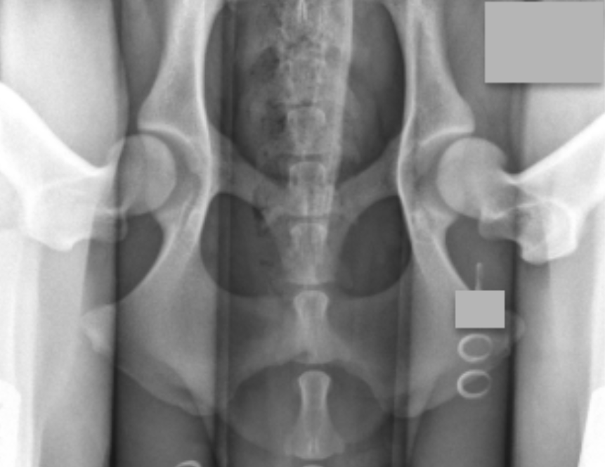
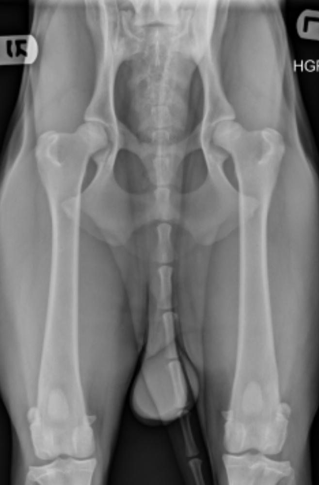
The obvious contrast in joint laxity between the distraction and hip-extended views demonstrates the fundamental difference between the two radiographs. The looser the joint on the distraction view, the greater is the chance that the hip will develop OA. The hip-extended view tends to mask true hip joint laxity because the joint capsule is wound up into a tightened orientation when the hips are extended. This explains why measurable joint laxity on the distraction view is always greater than the measurable laxity from the hip-extended view. In fact, distraction laxity can be up to 11 times greater, depending on the breed of dog under study.
The compression view is used to determine the “goodness of fit” of the femoral heads into the acetabula. In a hip with OA, the remodeling that occurs in the acetabulum and/or the femoral head will often result in an ill-fitting “ball” and “socket.”
To summarize, the PennHIP method:
Obtains OA readings from the standard hip-extended view.
Obtains hip joint congruity readings from the compression view.
Obtains quantitative measurements of hip joint laxity from the distraction view.