In-House Diagnostics
Element HT5™
Hematology Analyzer
CBC with a true 5-part differential, in under 60 seconds.
Proven Technology
Using flow cytometry, impedance, and colorimetry, the Element HT5™ provides fast and accurate results, including a true 5-part WBC differential.
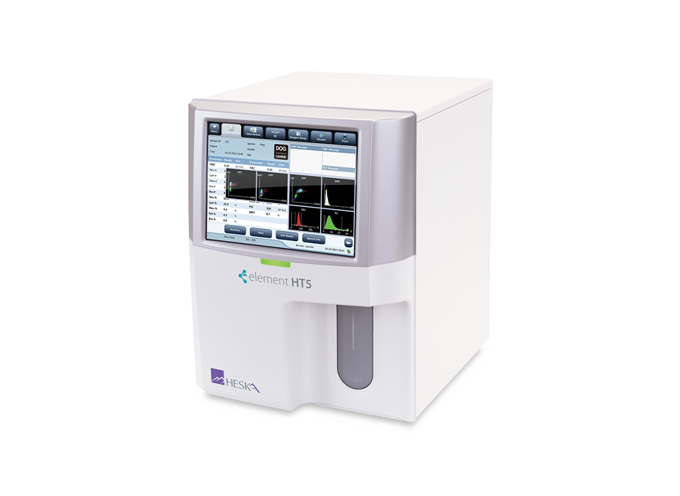
Touchscreen Interface
Color touchscreen with intuitive navigation simplifies data entry and test review through numeric results, histograms, and scatter plots.
Simple Data Management
Assign, unassign, or delete multiple items at once with easy web-style navigation links.
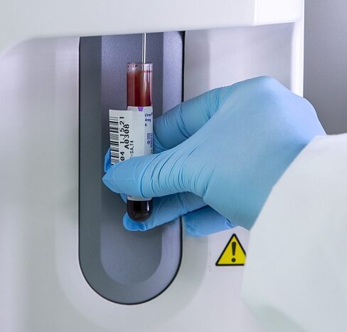
Easy Reagent Management
Barcode scanner and onboard menu functions simplify reagent management, with internal space-saving reagent storage.
Rapid Results
Results for 20 parameters in under 60 seconds.
Multiple Species Options
Dog, cat, horse, cow, ferret, goat, llama, camel, monkey, mouse, pig, rabbit, rat, sheep, giant panda, and red panda.
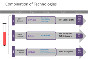
Laser + Impedance + Colorimetric Hematology = True 5-Part
Accuracy and Reproducibility
Excellent correlation for a population of animals measured on two different technologies.
Triple Angle Laser Scatter
Detects complexity and granularity of cell volumes.
Sample Pathology Messages
Intelligent information related to pathology present in the sample.
Technical Details and Downloads
Overview
Element HT5™
CBC with true 5-part differential, in only 60 seconds.
Three discrete measurement channels utilizing laser flow cytometry, impedance counts, and colormetric detection technologies provide an accurate CBC with true 5-part differential in only 60 seconds. The HT5™ can detect not only white blood cell volume, but also cellular compexity and granularity. Accurate, fast, and simple to use with large color touchscreen, intuitive navigation, simplified data entry, and easy reagent management. Fast enough for the busiest practices and cost effective for all.
Available Species:
Dog, cat, horse, cow, ferret, goat, llama, camel, monkey, mouse, pig, rabbit, rat, sheep, giant panda, and red panda.
Specs and Technology
Element HT5™ Measurement Methods Used:
- Electrical Impedance method for determining the RBC and PLT data
- Colorimetric Method for determining the HGB
- Flow Cytometry by Laser for determining the WBC data
Other parameter results are obtained via calculation.
Electrical Impedance Method
RBCs/PLTs are counted and sized with this method which is based on the measurement of changes in electrical resistance produced by a particle, in this case is a blood cell, suspended in a conductive diluent as it passes through an aperture of known dimensions. A pair of electrodes is submerged in the liquid on both sides of the aperture to create an electrical pathway. As each particle passes through the aperture, a transitory change in the resistance between the electrodes is produced. This change produces a measurable electrical pulse. The number of pulses generated represents the number of particles that passed through the aperture. The amplitude of each pulse is proportional to the volume of each particle.
Output includes numeric values, percentages plus histograms for RBC, WBC, and PLT plus scatter plots for analyzing cell volume, complexity, and granularity.
Flow Cytometry by Laser
After a predetermined volume of blood is aspirated and diluted by a certain amount of reagent, it is injected into the flow cell. Surrounded with sheath fluid (diluent), the blood cells pass through the center of the flow cell in a single column at a faster speed. When the blood cells suspended in the diluent pass through the flow cell, they are exposed to a laser beam.
The intensity of scatter light reflects the blood cell size and intracellular density. The low-angle scattered light reflects cell size, and the high-angle scattered light reflects intracellular density (nucleus size and density). The optical detector receives this scatter light and converts it into electrical pulses. Pulse data collected can be used to draw a three-dimensional distribution (scattergram).
Triple angle scatter detects cell volume, complexity, and granularity of cells.
Low Angle Scatter (LAS) detects cell volume
Mid Angle Scatter (MAS) detects cellular complexity
Wide Angle Scatter (WAS) detects cellular granularity
Multiple angle scatter plots aid to differentiate WBC differential.
Colorimetric Method
The WBC/HGB dilution is delivered to the HGB bath where it is bubble mixed with a certain amount of lyse, which converts hemoglobin to a hemoglobin complex that is measurable at 530 nm. An LED is mounted on one side of the bath and emits a beam of monochromatic light, whose central wavelength is 530 nm. The light passes through the sample and is then measured by an optical sensor that is mounted on the opposite side. The signal is amplified and the voltage is measured and compared to the blank reference reading (readings taken when there is only diluent in the bath), and the HGB is measured and calculated in the analyzer automatically.
Reticulocytes
Anemia is typically present in 5% to 10% of small animal samples. When anemia is detected, it’s useful to evaluate the blood for the presence of regeneration and RBC abnormalities that may help determine the cause of the anemia. Antech recommends assessing regeneration by examination of the stained blood film for polychromasia (or reticulocytes), while also looking for RBC abnormalities. The Element HT5 analyzer’s identification of an increase in RDW and/or MCV is evidence of probable reticulocytosis, but is best confirmed by slide examination.
Sample Pathology Messages
The Element HT5 system provides intelligent information messages related to pathology present in the sample, intended to indicate that diagnostic information found on the slide could supplement instrument results.
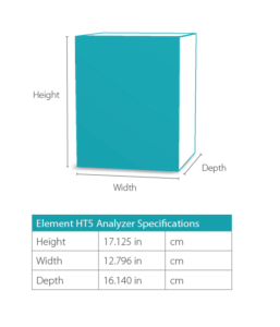
Dimensions
Interfaces
4 USB ports
1 Ethernet Port
Power Supply
Voltage Input power Frequency: Analyzer (100V-240V~)±10% 300 VA (50Hz/60Hz)±1Hz
Fuse
WARNING: Use specified fuse only. Fuse specification: 250V T3.15AH
EMC Description
Do not use this device in close proximity to sources of strong electromagnetic radiation (e.g., unshielded intentional RF sources), as these may interfere with the proper operation. This equipment complies with the emission and immunity requirements of the EN61326–1:2006 and EN61326–2–6:2006.
NOTE: It is the manufacturer’s responsibility to provide equipment electromagnetic compatibility information to the customer or user.
NOTE: It is the user’s responsibility to ensure that a compatible electromagnetic environment for the equipment can be maintained in order that the device will perform as intended.
Sound
Maximal sound: 65 dBA
Operating Environment
Optimal operating temperature: 50°F – 86°F
Optimal operating humidity: 20% – 85%
Atmospheric pressure: 70 kPa – 106 kPa
Storage Environment
Ambient temperature: 15°F – 104°F
Relative humidity: 10% – 90%
Atmospheric pressure: 50 kPa – 106 kPa
Running Environment
Ambient temperature: 50°F – 104°F
Relative humidity: 10% – 90%
Atmospheric pressure: 70 kPa – 106 kPa
NOTE: Be sure to use and store the analyzer in the specified environment
Resources
Instructions for Control File Download:
Right click on the control file.
Click on Save Link As…or Save Target As…depending on your browser.
Save to USB drive and then upload to Analyzer.
Hematology controls information for use
Hematology controls information for use (Canadian French)
Control Lot BC2607B, exp 09/10/2026
Control Lot BC2605B, exp 07/10/2026
Downloads
Antech™ Support
- Daily 6:00 a.m. to 5:00 p.m. MT
- Emergency support after hours
Consider these Antech™ solutions for the right healthcare, at the right time.
-
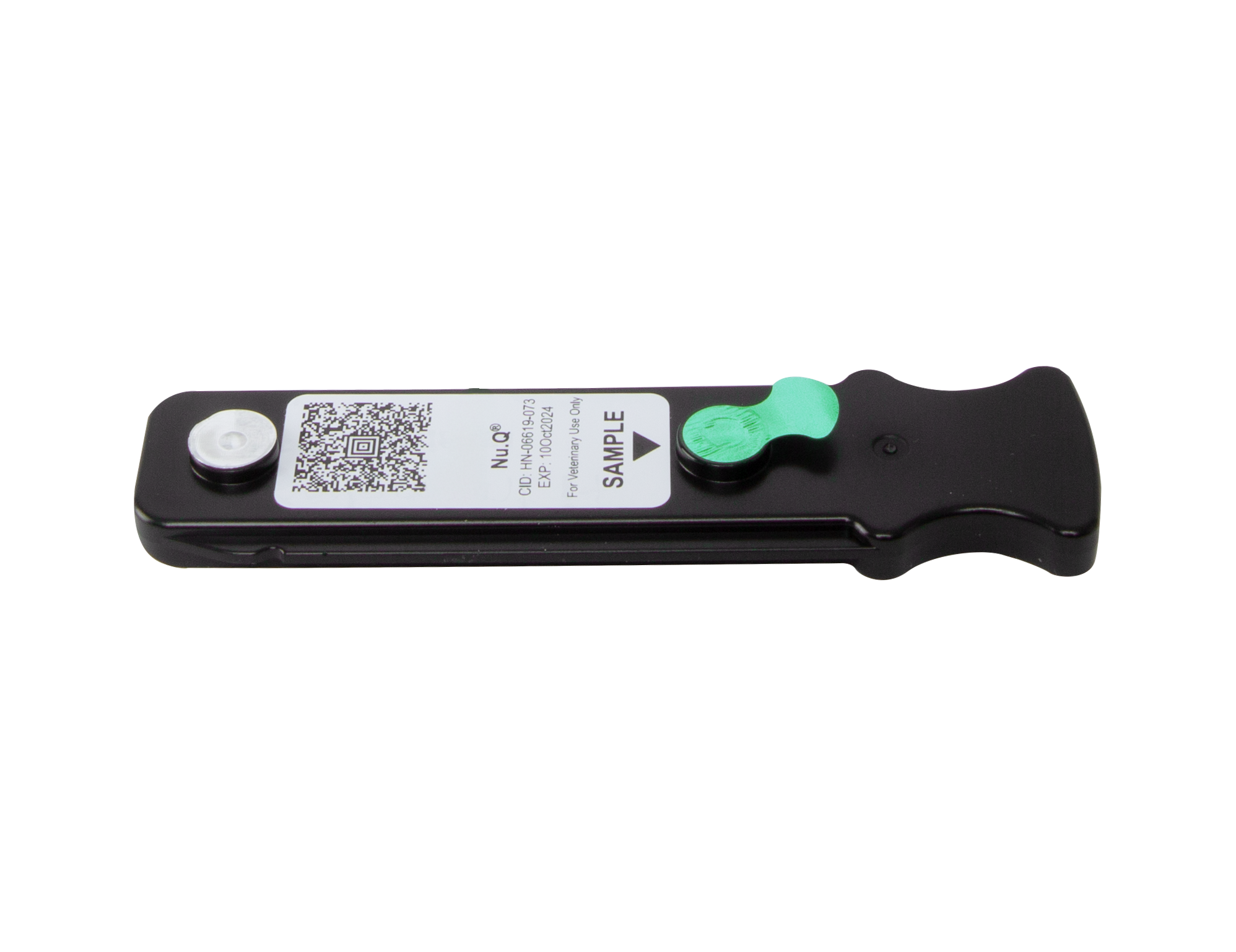
Nu.Q®
Empowering veterinary care teams with the industry’s only in-house canine cancer blood test.
Have questions about Element HT5™?
Complete the form and an Antech representative will reach out to you.